
The causes, compensatory reactions, mechanism of development of the injury. Examples of damaging action of physical, chemical and biological injuring factors. Category’s of the pathogenesis (causes and the effect of relationships, vicious circle, local and common, forms and function, nonspecific and specific).Ĭlassification of environmental factors, the role of ones in initiation of diseases. Concepts of development of a disease: Mechanical monocausalism, condicionalism, Freudism. The concept of pathological reaction, pathological process and pathological status. The structure of pathophysiological experiment and the tasks of each stage. Interrelation of pathophysiology with theoretical and clinical subjects. The subject, methods and tasks of pathophysiology. Generalized Type III hypersensitivity reaction at different site results in different diseases such as Glomerulonephritis (Kideny), vasculitis (arteries), Arthritis (synovial joints).Examination questions for the pathological physiology for the 3Įxamination questions for the pathological physiology for the 3 rd year students.The site may vary but accumulation of complexes occurs at site of blood filtration. The manifestation of serum sickness depends on the quantity of immune complex as well as overall site of deposition.

If antigens are in significantly excess compared to antibody, the immune complexes formed are smaller and soluble which are not phagocytosed by phagocytic cells leading to Type III hypersensitivity reaction.
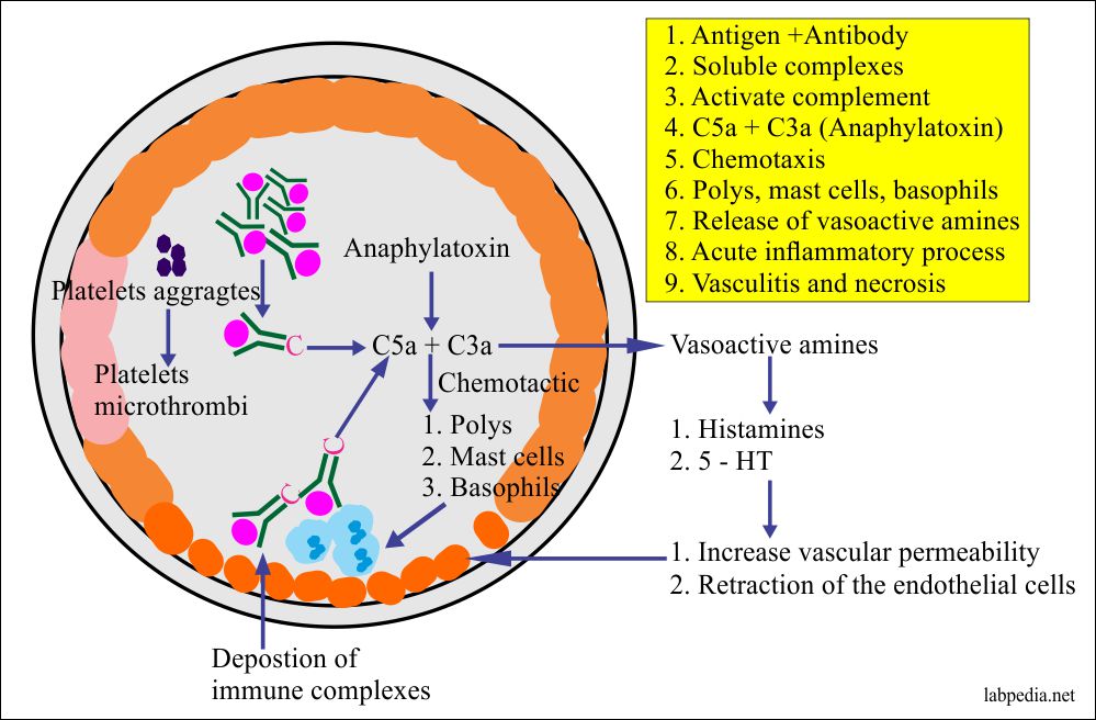
When large amount of antigen enter blood stream and bind to antibody, circulating immune complexes forms. Serum sickness is an example of generalized Type III hypersensitivity reaction. Generalized Type III hypersensitivity reaction: The severity of reaction can vary from mild swelling and redness to tissue necrosis.Ģ. As the reaction develops, localized tissue damage and vascular damage results in accumulation of fluids (edema) and RBCs (erythema) at the site of antigen entry. When antigen is injected or enters intradermally or subcutaneously, they bind with antibody to form localized immune complexes which mediate acute Arthus reaction within 4 to 8 hours. Acute Arthus reaction is an example of localized Type III hypersensitivity reaction. Localized Type III hypersensitivity reaction: Types of Type III hypersensitivity reaction: 1. Furthermore complement proteins can also contribute to tissue destruction. The lytic enzymes cause tissue damage surrounding of immune complex deposits, resulting hypersensitivity reaction. The neutrophils attempt to phagocytose the immune complex but phagocytosis is not possible because immune complexes are deposited on basement membrane, so the neutrophil releases lytic enzymes to destroy immune complex. Neutrophil binds to C3b coated immune complex by means of type I complement receptor which is specific for C3b. C3b acts as opsonin by binding with immune complex. C5a, C3a and C5b67 also acts as chemotatic factors for neutrophils, So it attracts neutrophils at the site of immune complex deposition. This facilitates deposition of immune complexes on wall of blood vessel. Degranulation of mast cell releases histamine which increases vascular permeability of blood capillaries. C3a and C5a are lymphotoxin (anaphylotoxin) that causes localized mast cell degranulation. Type III hypersensitivity reaction develops when immune complex activates C3a and C5a components of complement system. Deposition of immune complexes initiates reaction resulting in damage of surrounding tissue and cause inflammation. Type III hypersensitivity reaction is characterized by deposition of immune complexes on various tissues such as wall of blood vessels, glomerular basement membrane of kidney, synovial membrane of joints and choroid plexus of brain. If immune complexes are not removed from blood, they accumulate on wall of blood vessels and on tissue where filtration of blood and plasma occurs such as glomerular membrane, synovial membrane of joints etc. In some cases large amount of immune complexes are formed and deposited on various body parts and leads to tissue damage resulting in Type III hypersensitivity reaction. Complement system is also needed for removal of immune complexes from blood to spleen. Most other immune complexes are first carried by blood to spleen where they are destroyed by macrophages. Some immune complexes are removed by phagocytic action of phagocytic cells in blood. In most of the cases, these immune complexes are removed from blood circulation. 
The reaction of antibody with antigen generates immune complex.Type III hypersensitivity reaction: factors causing immune complex formation, mechanism and types


 0 kommentar(er)
